Do They Break Your Ribs for Open Heart Surgery? Sternotomy Explained
 Nov, 13 2026
Sternotomy Recovery & Safety Checker
Enter your days since surgery to see where you are in the healing process and what activities are generally safe.
Activity Safety Simulator
Select an activity to see if it is generally recommended at your current recovery stage.
You are sitting in the pre-op holding area. The nurse is checking your vitals. You look at the chest X-ray on the monitor and a sudden, primal fear hits you: Are they going to break my ribs?
It’s a question that haunts almost everyone facing open heart surgery. It sounds brutal-like something out of a medieval torture chamber rather than a modern hospital. But here is the short answer: No, they don’t break your ribs. Not in the way you’re imagining. There is no hammer, no accidental snap, and no jagged bone shards. What actually happens is a controlled separation of the breastbone, a procedure called a sternotomy. Understanding the difference between "breaking" and "separating" can take a massive weight off your shoulders before you even walk into the theatre.
The Myth of the Broken Rib
Let’s clear up the confusion immediately. When people hear "open heart," they picture a surgeon cracking the chest like an eggshell. That’s not how it works. The human chest cavity is protected by the rib cage, which is rigid. To get to the heart, surgeons need access. In traditional open heart surgery, they don't go through the ribs themselves; they go down the middle, through the sternum, also known as the breastbone.
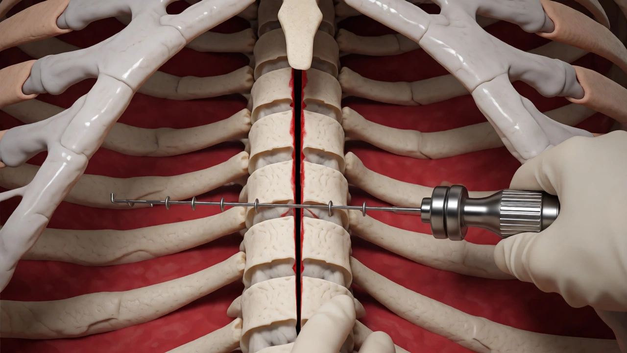
The sternum is a flat bone, not a long tubular bone like your femur or radius. It is held together by cartilage and fibrous tissue. During a median sternotomy, the surgeon uses a specialized oscillating saw to cut vertically through the center of this bone. Think of it less like breaking a stick and more like splitting a log along its grain. Once cut, the two halves of the sternum are gently spread apart using a retractor. This creates a window to the heart without shattering the structural integrity of the surrounding rib cage.
| Feature | Sternotomy (Surgical Cut) | Rib Fracture (Trauma/Break) |
|---|---|---|
| Cause | Controlled surgical incision with a saw | Blunt force trauma or severe coughing |
| Bone Integrity | Clean cut; ends remain aligned | Displaced fragments; potential puncture risk |
| Healing Process | Bone knit back together with wires | Natural healing; may heal with deformity |
| Pain Type | Deep ache, pressure when moving torso | Sharp, stabbing pain with breathing |
How a Sternotomy Actually Works
So, if they aren’t breaking ribs, what exactly are they doing? The process is highly technical and relies on precision tools. After you are under general anesthesia, the surgeon makes an incision down the center of your chest. Then comes the oscillating saw. This isn’t a circular saw like you’d use in a workshop. It vibrates rapidly back and forth, cutting only hard bone while leaving softer tissues (like the skin above and the heart below) relatively untouched. It’s surprisingly quiet compared to a power tool, but you’ll hear the sound if you’ve ever been in the room during induction.
Once the cut is complete, the surgeon inserts a device called a sternal retractor. This looks a bit like a giant metal clamp. It slowly spreads the two halves of the sternum apart, creating about 4-6 inches of space. This gap allows the heart-lung machine and the surgeon’s hands to work freely. Crucially, the ribs attached to the sternum move slightly outward with this spread, but they do not break. They flex. Human bones have a certain degree of elasticity, especially in younger patients, though this decreases with age.
At the end of the operation, once the heart repair is done, the surgeon doesn’t just glue the bone back. They use stainless steel wires to lace the two halves of the sternum together, similar to lacing a shoe. These wires hold the bone firmly in place while it heals. Over the next 6 to 8 weeks, new bone tissue grows across the cut line, fusing the sternum back into a single solid unit. The wires usually stay in forever-they’re small, inert, and rarely cause issues. If you ever get a CT scan or MRI later in life, radiologists will see them, but they won’t set off airport security alarms.
Why Do We Still Use This Method?
If it sounds complicated, why not just go between the ribs? You might ask. For many procedures, we actually do. But for complex operations like coronary artery bypass grafting (CABG) or valve replacements, the sternotomy offers the best visibility. The heart sits centrally in the chest. Going through the middle gives the surgeon direct access to all four chambers and the major vessels without having to navigate around the dense mesh of ribs and intercostal muscles.
There is also safety to consider. If a complication arises during surgery-say, unexpected bleeding-the surgeon needs immediate, wide access to control it. A sternotomy provides that rapid exit strategy. Minimally invasive techniques are great, but they require specific anatomical conditions and skilled teams. For emergency surgeries or complex multi-vessel disease, the old-school sternotomy remains the gold standard because it is fast, reliable, and familiar to every cardiac surgeon in the world.
Recovery: The "Broken Bone" Sensation
Here is where the myth persists. Even though the ribs weren’t broken, the recovery feels a lot like a fracture. Why? Because you have effectively severed a major load-bearing bone in your torso. Every time you twist, lift, or breathe deeply, you engage the pectoral muscles and the intercostal muscles, which pull on the healing sternum.
- Weeks 1-2: Pain is highest here. You’ll likely be on strong analgesics. Moving from bed to chair feels monumental. Coughing is terrifying because it jars the chest. Hospitals provide pillows to hug against your chest when you cough-a technique called "splinting" that reduces pain significantly.
- Weeks 3-6: The acute pain subsides, replaced by a deep, dull ache. You can start walking outside. Driving is usually off-limits until you can check your blind spot without wincing, typically around week 4-6.
- Months 2-3: The bone is knitting. Most patients report feeling "normal" again, though heavy lifting (over 10 lbs) is still restricted. You might feel clicking sensations in the chest; this is often muscle tension or minor wire movement, not the bone breaking.
Patients often worry that their sternum will pop open if they sneeze or roll over in bed. It’s rare, but possible. This condition is called sternal dehiscence. It happens more often in smokers, diabetics, or those who ignore lifting restrictions. If you feel a distinct "clunk" or sudden sharp pain followed by instability, call your surgeon. But for the vast majority, the wires do their job perfectly.

Minimally Invasive Alternatives
Not every heart surgery requires cracking the chest. Medicine has advanced, and depending on your specific diagnosis, you might qualify for less invasive options. These approaches avoid the sternotomy entirely, meaning no bone cutting at all.
| Procedure | Incision Location | Bone Impact | Best For |
|---|---|---|---|
| Median Sternotomy | Center of chest | Sternum split | Complex CABG, multiple valves |
| Thoracotomy | Side of chest (between ribs) | Ribs spread, not cut | Lung biopsy, some mitral repairs |
| Mini-Thoracotomy | Small side incision | Minimal rib impact | Single vessel bypass, TAVR access |
| Robotic Surgery | Several small ports | No bone cutting | Mitral valve repair, ASD closure |
For example, Transcatheter Aortic Valve Replacement (TAVR) involves inserting a new valve through a catheter in your groin artery. No chest incision, no bone cutting, and you could potentially go home the next day. Similarly, robotic-assisted mitral valve repair uses three tiny holes instead of a large central scar. However, these aren’t available for everyone. If you need bypasses on three different arteries, the robot’s dexterity might not match the speed and visibility of an open sternotomy. It’s a trade-off between cosmetic appeal/speed of recovery and surgical complexity.
Tips for Protecting Your Chest Post-Surgery
Since the sternum takes about 6-8 weeks to heal fully, your behavior during this time dictates your comfort level. Here are practical rules to live by:
- The 10-Pound Rule: Don’t lift anything heavier than a gallon of milk with both arms extended. This includes grandchildren, groceries, and gym weights. Use your legs to lift, keeping objects close to your body.
- Push Up Technique: Never push yourself up from a chair using your arms alone. This puts immense strain on the chest. Instead, lean forward, shift your weight to your hips, and stand up using leg strength. Or better yet, slide your hands up your thighs.
- Log Roll: Get in and out of bed by rolling onto your side first, then pushing up with one arm while swinging your legs over the edge. Twisting directly from lying on your back pulls the sternum apart.
- Hygiene Hack: Showering is tricky early on. Use a handheld showerhead so you don’t have to reach overhead or twist to wash your back. Many hospitals recommend sponge baths for the first week to keep the incision dry.
Remember, the goal isn’t to immobilize yourself completely. Walking is encouraged immediately after discharge. Movement prevents blood clots and pneumonia. Just move smartly. Listen to your body. If a movement causes sharp chest pain, stop. Discomfort is normal; agony is not.
Does the sternum grow back together?
Yes. The sternum is a living bone. After being wired together, osteoblast cells create new bone matrix across the surgical cut. Within 6 to 8 weeks, the bone fuses into a solid structure again. The steel wires remain inside to provide permanent stability, but the biological connection is restored.
Will I have a visible scar?
You will have a vertical scar down the center of your chest, typically 6-8 inches long. Initially, it may be red and raised (hypertrophic), but it fades over 12-18 months to a pale white line. Some patients develop keloids, especially if they have darker skin tones, but silicone sheets and massage can help minimize appearance.
Can I exercise after heart surgery?
Absolutely, and you should. Cardiac rehabilitation programs are designed specifically for post-surgery patients. You start with low-intensity walking and gradually progress to cycling, swimming, and resistance training. By month 3-4, most patients return to full activity levels, including jogging and weightlifting, provided their heart health permits it.
What if I feel clicking in my chest?
Clicking or popping sensations are common during the first few months. It is often caused by the sternum shifting slightly within the wires or by tight chest wall muscles stretching. As long as there is no significant pain, swelling, or redness, it is usually harmless. Mention it to your doctor at your follow-up appointment for peace of mind.
Is minimally invasive surgery always better?
Not necessarily. While smaller incisions mean faster recovery and less pain, they offer limited access. For complex cases involving multiple blockages or combined procedures (e.g., valve replacement plus bypass), the superior visibility and safety of a sternotomy outweigh the cosmetic benefits of keyhole surgery. Your cardiologist will weigh risks versus benefits based on your anatomy.